Microscope images of stained sperm
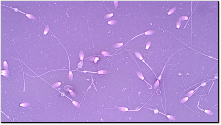

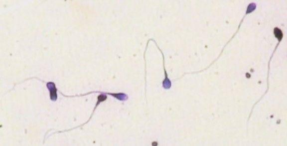

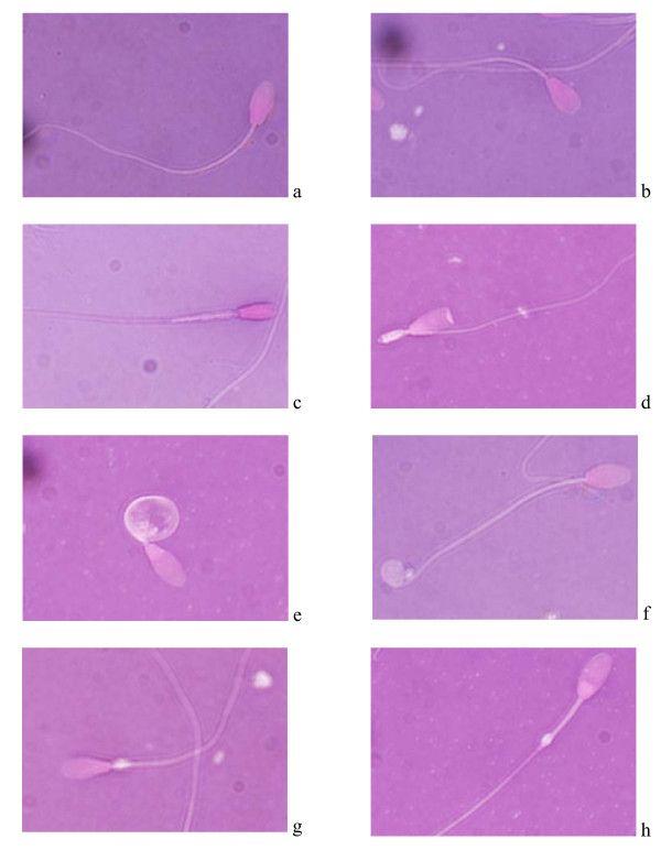
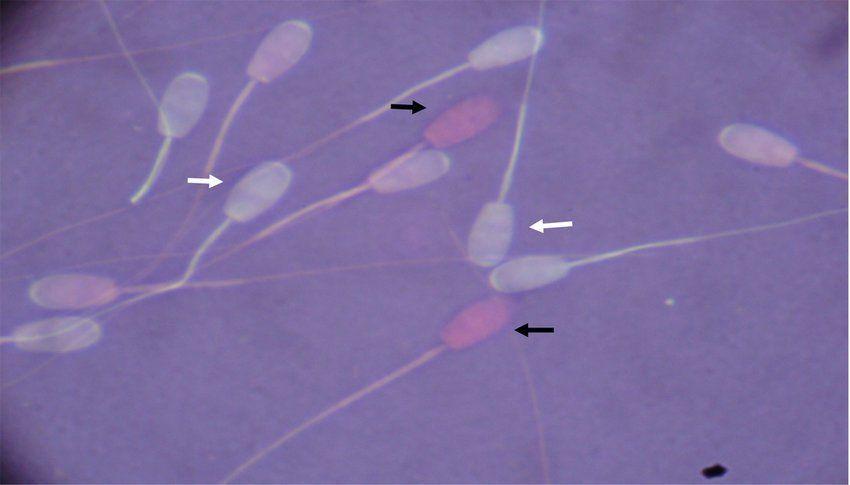
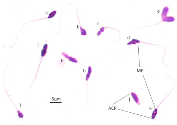
13 Feb The morphology seen with the microscope is not the true morphology of a living spermatozoon, but an image we create. This image comprises a staining) are of high quality when assessing sperm morphology. Even small.
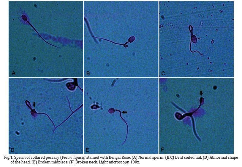
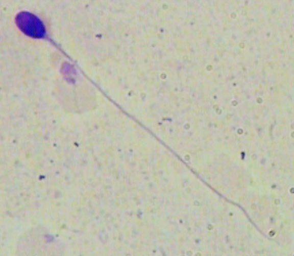
Sperm's tail identification and discrimination in microscopic images of stained human semen smear. Abstract: Correct estimating of a man's fertility potential has .




Kay Age: 25. ?Fetish friendly ????Premium Public Records for name Larry G Dick found in this find people section originate from public directories available on the internet to their subscribers.Clean and beautiful sex partherIf you are looking for some.
the microscope. How to interpret boar sperm morphology when PHOTO 1: Seen through a phase-contrast microscope, this grosin staining technique.
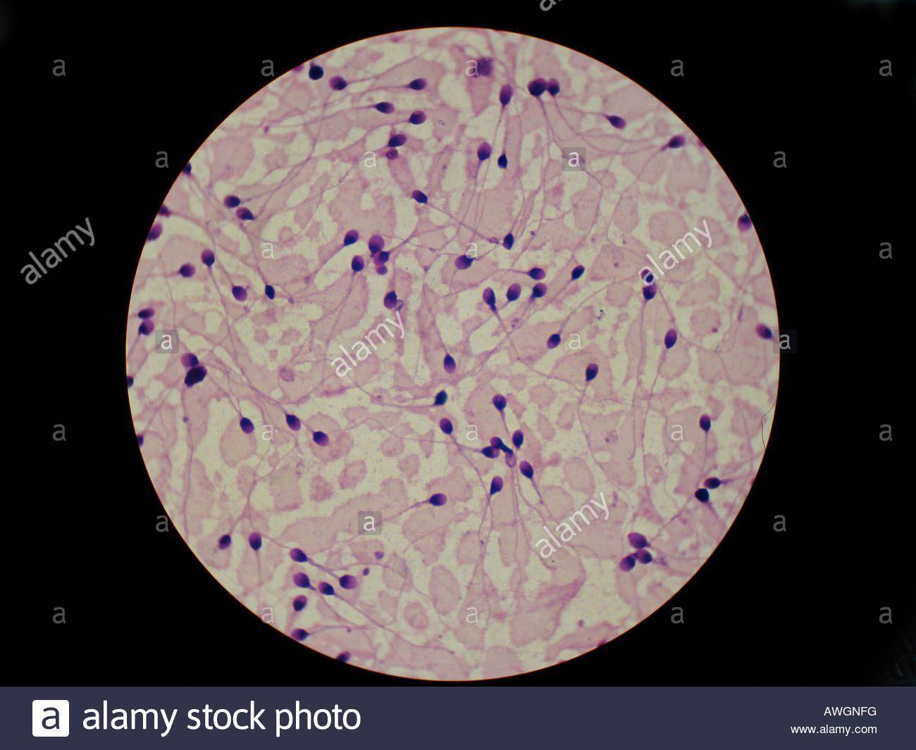
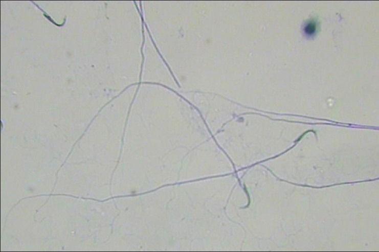
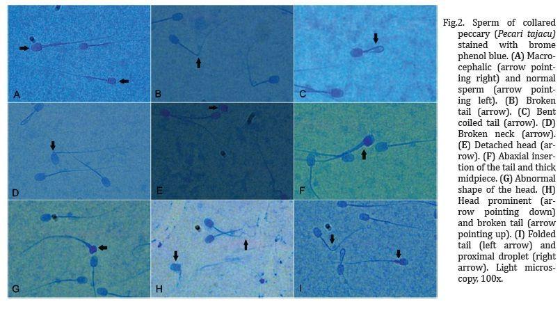
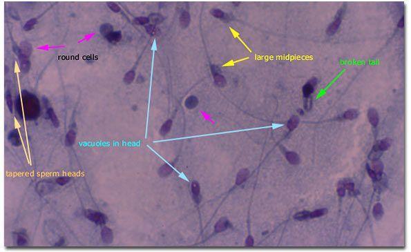
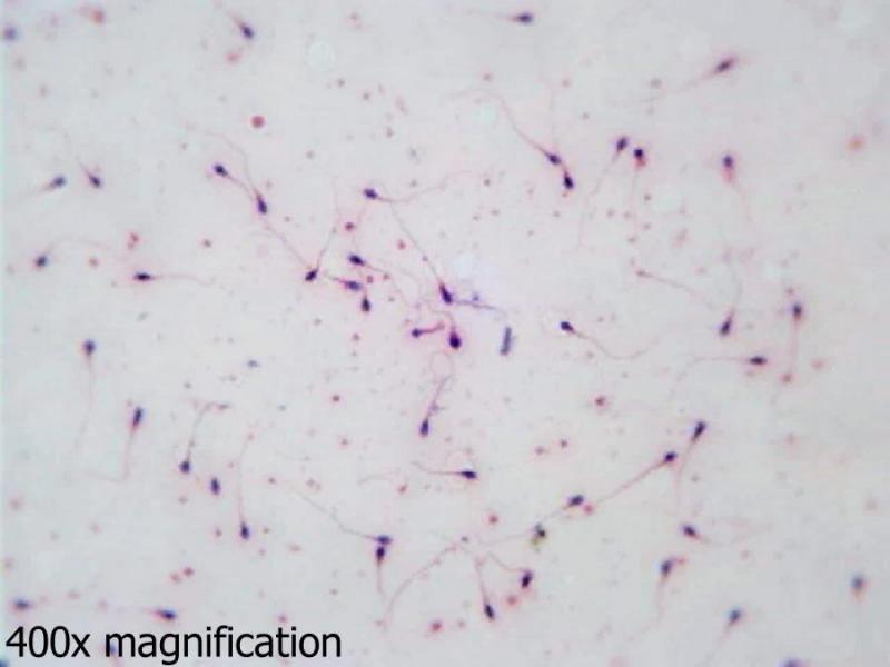
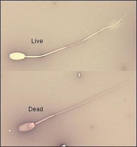
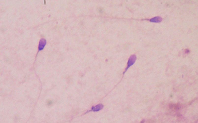
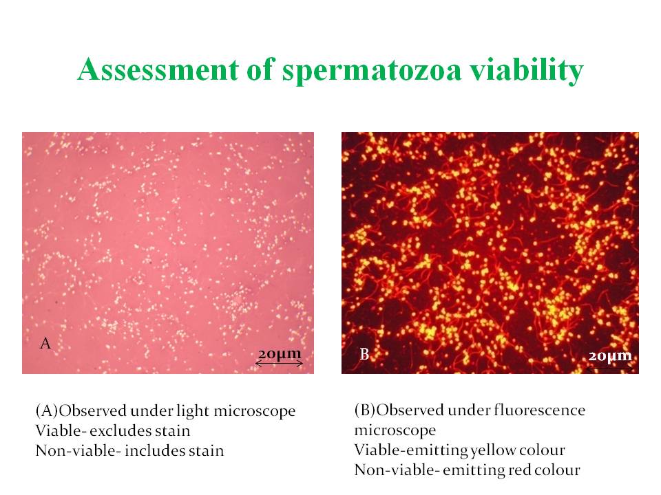
Figure 3 : Sperm viability and acrosome reaction analysis.

Scientific Reports
Description:This abnormal sperm has one X and 18 chromosomes, but two 13 and 21 chromosomes. All semen samples were analyzed within a day after collection. The existence of a positive correlation between fertility and sperm head morphometry was demonstrated in swine Villalobos et al. These photos show human embryos that are 3 days old. Each electrical stimulus lasted for 3s with intermittent breaks of 2s.


User Comments 2
Post a comment
Comment: